


Historical background:
The term ‘Angina’ is derived from Latin and Greek word for choking or strangling. Placing both into context, Ludwig’s angina refers to the feeling of strangling and choking, secondary to obstruction of the airway, which is the most serious complication of this syndrome.[1] It is named after a German physician, Wilhelm Frederick von Ludwig, who first described this condition in 1836.[2] Ludwig’s angina is hazardous because it can result in oedema and distortion, which obstructs the airway and makes breathing difficult due to the tongue’s elevation and posterior displacement.
In 1939, Dr.Manual Grodinsky developed the following diagnostic criteria for the diagnosis of Ludwig’s angina.[3]
‘There must be cellulitis, not an abscess, of the submandibular space that never involves only one space and usually is bilateral; produces gangrene with serosanguineous, putrid infiltration but very little frank pus; involves connective tissue, fascia, and muscles but not glandular structures; and is spread by continuity and not by lymphatics’.
Clinical presentation
A slight infection sets off Ludwig’s angina, which then causes induration of the upper neck, discomfort, trismus, and tongue elevation. Ludwig’s angina can be triggered by an initial minor infection usually in the 2nd and 3rd Mandibular molar teeth[4].
Ludwig’s angina can present with a variety of signs and symptoms, including tachycardia, fever, malaise.
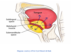
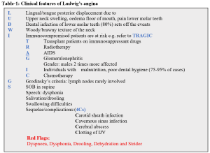
The clinical presentation depends on the severity but it could be described as LUDWIG’S angina as outlined in the table-1 below.
Table-1: Clinical features of Ludwig’s angina
Differential Diagnosis
- Angioedema
- Pemphigus
- Retropharyngeal abscess
- Lemierre’s syndrome is a rare and dangerous bacterial (Fusobacterium necrophorum) complication of sore throat or other head/neck infection that causes blood clots in the IJV and spreads infected clots through blood stream.
- Vincent’s angina which is the inflammation of pharynx and tonsils caused by two types of bacteria i.e. Fusobaterium and Spirochaetes (Borrelia and Spirillum). It is named after a French physician, Jean H Vincent. If the same organisms infect the gums causing ulcerative gingivitis, it is called Trench mouth as it was commonly seen in soldiers in the battle field (trench) during the 1st World War.
Diagnosis of Ludwig’s Angina
Blood culture takes up to 7 days
Imaging: contrast-enhanced scans of the neck, chest and head are gold standards for visualising the infected clot(thrombophlebitis) in the jugular vein and spotting whether the infection has spread to lungs, brain etc.
Treatment:
- IV antibiotics for 3 weeks targeting anaerobes: co-amoxiclav IV and Metronidazole IV
- Dexamethasone 10 mg IV regularly to reduce soft tissue oedema
- Airway secured before obstruction happens
- Surgical drainage of serosanguinous fluid +drains needed
- Nutritional support
Anaesthetic management of Ludwig’s angina
Ludwig’s angina may present with bilateral cervical swelling, dysphagia, drooling, neck tenderness, elevation, posterior displacement and swelling of the tongue, restricted neck movements, trismus, dyspnoea, and stridor, all of which can complicate airway management [5]. Therefore, it is essential to thoroughly assess the patient preoperatively to identify features that may cause difficulties with mask ventilation, direct laryngoscopy, and intubation. Alternative methods of securing airway should be considered and readily accessible, along with a clear plan for their use if mask ventilation or intubation proves impossible [6]
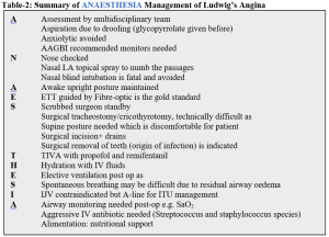
Conventional orotracheal intubation under general anaesthesia in patients with Ludwig’s angina is challenging. The distorted airway anatomy, tissue immobility, and limited access to the mouth make orotracheal intubation with conventional laryngoscopy difficult or impossible. The anaesthetic management is briefly outlined in table-2.
Table-2: Summary of ANAESTHESIA Management of Ludwig’s Angina
Conclusion
Ludwig’s angina, although uncommon, remains a potentially life-threatening condition because of the risk of impending airway obstruction.
Effective treatment is based on early recognition of the clinical process, with the appropriate use of parenteral antibiotics, securing the airway, and formal surgical drainage of the infection. Awake (or sedation with dexmedetomidine) fibreoptic intubation under topical anaesthesia may be the preferred method to secure the airway [7,8,9,10,11]. When fibreoptic bronchoscopy is not feasible, not available, or has failed, an elective awake cricothyrotomy and tracheostomy are the options. The patient needs to be nursed in critical care unit or HDU.
Conflict of Interest: None declared
Authors and Contributors:
Dr. Sher Mohammad1 Dr. Syed Murad Ali Shah2 Dr. Ubaid Urehman3 Dr. Izhar Ullah Khan4 Dr. Nazeer Ahmed5 Dr. Liaqat Ali6 Dr. Mohammad Ahmad Saroosh Saleem7 Dr. Habib Ben Aslam8
1.Consultant Anaesthetist(retired), STH NHS FT Sheffield
2.Proessor of Oral and Maxillofacial Surgery, Sardar Begum Dental College, Peshawar
3.Postgraduate Trainee, Oral and Maxillofacial Surgery, Rehman College of Dentistry. Peshawar
4.Clinical attachment c/o Prof. Syed Murad Ali Shah, Sardar Begum Dental College, Peshawar
5.Consultant Anaesthetist, Hamad General Hospital, Doha Qatar
6.Professor of Anaesthesia, Services Institute of Medical Sciences, Lahore
7.Senior Registrar Anaesthetics, Shalimar Hospital Lahore
8.Consultant Emergency Medicine, Great Western Hospital, Swindon SN3 6BB
References
- Costain N, Marrie TJ (February 2011). “Ludwig’s Angina”. The American Journal of Medicine. 124 (2): 115–7. doi:10.1016/j.amjmed.2010.08.004. PMID 20961522.
- Murphy SC (October 1996). “The person behind the eponym: Wilhelm Frederick von Ludwig (1790-1865)”. Journal of Oral Pathology & Medicine. 25 (9): 513–5. doi:10.1111/j.1600-0714. 1996. tb00307.x. PMID 8959561.
- Patterson HC, Kelly JH, Strome M. Ludwig’s angina: an update. Laryngoscope. 1982;92:370-378.
- Tschiassny K: Ludwig’s angina: an anatomic study of the role of the lower molar teeth in its pathogenesis . Arch Otolaryngol. 1943, 38:485-496. 10.1001/archotol.1943.00670040504010
- Ovassapian A, Warner DO: Principles of airway management, Third Edition. Anesthesiology. 2004, 101:1255. 10.1097/00000542-200411000-00049
- Cattano D, Killoran PV, Iannucci D, et al.: Anticipation of the difficult airway: preoperative airway assessment, an educational and quality improvement tool. Br J Anaesth. 2013, 111:276-285. 10.1093/bja/aet029
- Suehara AB, Gonçalves AJ, Alcadipani FA, Kavabata NK, Menezes MB. Deep neck infection-analysis of 80 cases. Braz J Otorhinolaryngol. 2008;74(2):253-9.2.
- Parhiscar A, Har-El G. Deep neck abscess: a retrospective review of 210 cases. Ann Oto Rhinol Laryngol 2001;110(11):1051-4
- Ovassapian A, Krejcie TC, Yelich SJ, Dykes MH. Awake fibreoptic intubation in the patient at high risk of aspiration. Br J Anaesth. 1989;62(1):13-6.4.
- Mazlan MZ, Hassan SK, Mukmin LA, Hassan MH, Abiddin HZ, Mohamad I, Nadarajan C, Muthusamy R, Eu CS. Novel usage of dexmetomidine in a paediatric patient with giant tongue haemangioma. Mal J Med Health Sci 2016;12(2):46-8.5.
- Furst IM, Ersil P, Caminiti M. A rare complication of tooth abscess-Ludwig’s angina and mediastinitis. J Can Dent Assoc. 2001;67(6):324-9
